Translate this page into:
Subcutaneous fat necrosis of the newborn with severe hypercalcemia in an infant

-
Received: ,
Accepted: ,
How to cite this article: George A, Yadav J, Dayal D. Subcutaneous fat necrosis of the newborn with severe hypercalcemia in an infant. J Pediatr Endocrinol Diabetes 2023;3:82-3.
Abstract
A 7-week-old male infant presented with erythematous skin nodules and hypercalcemia. The baby was diagnosed with subcutaneous fat necrosis of the newborn. The baby was managed with analgesics and various calcium-lowering measures.
Keywords
Hypercalcemia
1-alpha-hydroxylase
neonate
CASE SUMMARY
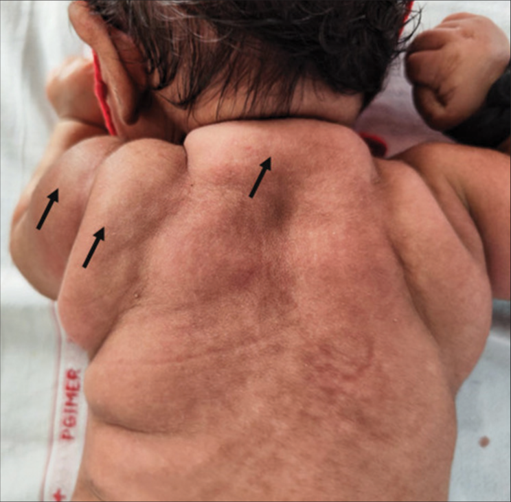
A 7-week-old boy presented with distinct erythematous and indurated and tender nodules on the upper trunk and limbs noted from birth, along with vomiting from the 4th week of life [Figure 1]. He was born at term to a primigravida mother by an emergency cesarean section performed in view of antenatal oligohydramnios, with subsequent birth asphyxia necessitating 7-day ventilation. Episodes of vomiting and constipation prompted assessment, revealing hypercalcemia (serum calcium 22 mg/dL), leading to a referral for further management. The evaluation showed low vitamin D (3 ng/mL, normal >20), elevated 1,25-dihydroxyvitamin D (>200 pg/mL, normal 34.8–88.8), and suppressed parathyroid hormone (1.14 pg/mL, normal 15–65). A diagnosis of subcutaneous fat necrosis (SCFN) was established in the context of characteristic skin lesions, hypercalcemia, hypercalciuria, and nephrocalcinosis. The baby was managed with intravenous hydration, furosemide, subcutaneous calcitonin, and bisphosphonates (pamidronate 1 mg/kg). On follow-up, the lesions gradually resolved, and his calcium levels (10 mg/dL) returned to normal.

- Multiple areas of fat necrosis seen on the back of the child (black arrows).
DISCUSSION
SCFN is a rare panniculitis that harbors 1-alpha-hydroxylase activity. Pre-eclampsia, diabetes mellitus in the mother, cord around the neck, hypothyroidism, cord prolapse, birth asphyxia, and therapeutic hypothermia are common risk factors. It presents with painful erythematous lesions, hypoglycemia, thrombocytopenia, and hypercalcemia. Management of pain and hypercalcemia with analgesics, hyperhydration, diuretics, calcitonin, and bisphosphonates is the crux of care.[1,2]
Declaration of patient consent
The authors certify that they have obtained all appropriate patient consent.
Conflicts of interest
There are no conflicts of interest.
Use of artificial intelligence (AI)-assisted technology for manuscript preparation
The authors confirm that there was no use of artificial intelligence (AI)-assisted technology for assisting in the writing or editing of the manuscript and no images were manipulated using AI.
Financial support and sponsorship
Nil.
References
- Subcutaneous fat necrosis in newborns: A systematic literature review of case reports and model of pathophysiology. Mol Cell Pediatr. 2022;9:18.
- [CrossRef] [PubMed] [Google Scholar]
- Subcutaneous fat necrosis of the newborn and associated hypercalcemia: A systematic review of the literature. Pediatr Dermatol. 2019;36:24-30.
- [CrossRef] [PubMed] [Google Scholar]


